|
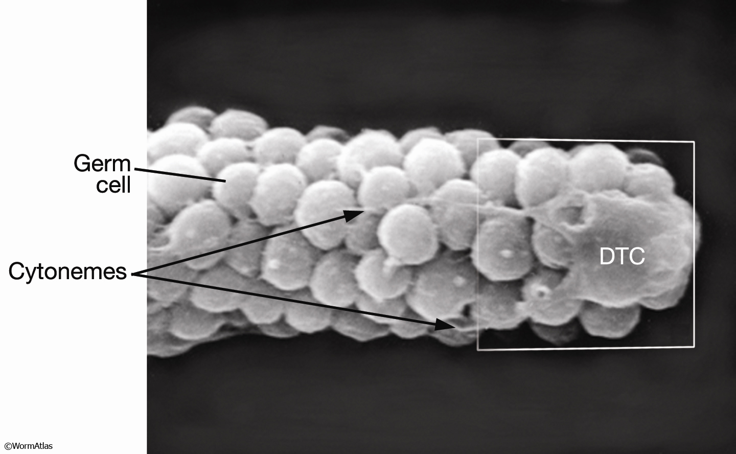
EMSEMDissectionFIG 1: Scanning electron micrograph of the DTC from dissected gonad.
Lateral view. Distal-most end of an adult gonad (dissected away from the rest of the body). Long thin trailing processes of the DTC extend (leftward) across the unsheathed distal arm of the mitotic gonad. A bulge on the leading edge of the DTC (far right) may be the nucleus being pushed forward. (Image source: L. Hoffman and D. Greenstein, photo #003.)
Click on picture for full resolution image.
|